
Skin cancer remains the most common cancer worldwide, and melanoma is the most dangerous type. According to the World Health Organization, around ~325,000 new cases of melanoma are diagnosed globally each year, and early detection is the single most effective way to save lives. Skin Mole mapping is one of the most practical and advanced methods for spotting melanoma and other skin cancers early.
For individuals at higher risk—such as those with fair skin, a history of sunburns, or many atypical moles—mole mapping provides a level of accuracy and reassurance that regular visual checks simply cannot.
Unlike a quick glance in the mirror or even a standard skin exam, mole mapping uses specialized photography and digital tracking tools to create a detailed record of your skin. Each mole and mark is documented, measured, and stored, allowing dermatologists to compare images over time.
Subtle changes that could otherwise be missed become visible, often months or years before they might be detected in routine checks. This early intervention can dramatically reduce the chances of melanoma spreading, and in most cases, treatment is far simpler and less invasive when cancers are caught early.

How Mole Mapping Works in Practice?
The process of mole mapping begins with an initial consultation. At this stage, the dermatologist carefully examines the patient’s skin and medical history to assess risk factors for melanoma and other skin cancers.
If mole mapping is considered appropriate, a series of high-resolution digital photographs is taken to establish a baseline record of the patient’s entire skin surface. These images include areas that are difficult for patients to check themselves, such as the scalp, back, and behind the ears. Each mole or pigmented lesion is documented, and specialized software stores the images in the patient’s digital profile.
During follow-up visits, the dermatologist compares the new photographs with the baseline images. This enables even the smallest changes in size, shape, or pigmentation to be detected with millimeter precision. Such accuracy is not possible through memory or routine self-examination. Research shows that over half of melanomas arise as new lesions rather than from pre-existing moles, which highlights the importance of monitoring both established and newly developed spots.
Mole mapping is particularly useful for patients with numerous moles, atypical mole patterns, or a family history of melanoma. Without photographic records, it can be extremely difficult to notice subtle changes over time. By providing an objective and systematic comparison, mole mapping helps dermatologists make earlier and more reliable diagnoses, ultimately improving patient outcomes.
Why Mole Mapping is More Reliable Than Self-Checks?
Self-exams are important, but they have limitations. Many people don’t notice slow changes, and areas like the back or scalp are nearly impossible to monitor alone. Dermatologists use mole mapping to reduce these blind spots. Here is a clear comparison:
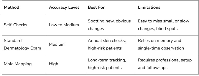
By using mole mapping, subtle changes that may signal the very first stage of melanoma are caught before they pose serious danger. This gives patients more confidence and doctors more data to make precise decisions.
Important Tips for Patients Considering Mole Mapping
Choose a qualified clinic: Not all skin clinics offer advanced mole mapping. Ensure your provider uses high-resolution digital imaging and has dermatologists who specialise in skin cancer detection.
Schedule regular follow-ups: Mapping is most effective when repeated at consistent intervals. Most patients benefit from annual comparisons, while higher-risk individuals may need check-ups every 6 months.
Don’t ignore self-checks: Even with mapping, you should still keep an eye on your skin at home. If you notice a new or rapidly changing spot, don’t wait for your next appointment.
Know your risk level: People with a family history of melanoma, fair skin, or a high number of moles are at greater risk. Mapping can provide life-saving reassurance in these cases.
Combine with sun safety: Mole mapping is not a substitute for prevention. Daily sunscreen use, protective clothing, and avoiding peak UV hours remain essential steps.
Benefits Beyond Melanoma Prevention
While melanoma is the primary concern, mole mapping can also help in detecting other skin cancers, including basal cell carcinoma and squamous cell carcinoma. These are more common than melanoma and often appear in sun-exposed areas. By identifying unusual lesions early, mole mapping can help ensure treatment is quick and straightforward, reducing scarring and recovery time.
Additionally, mole mapping provides peace of mind. For many patients, anxiety about whether a mole has changed is as distressing as the actual health risks. Having a professional, photographic record removes the guesswork and provides clear evidence over time. Parents often choose mole mapping for children with numerous birthmarks or moles, ensuring early protection as they grow.
Mole Mapping and Skin Cancer Prevention Questions
Is mole mapping painful?
No. The process is completely non-invasive and involves only photography. There is no discomfort or recovery time.
How often should I do mole mapping?
Most people benefit from annual mapping. If you are high-risk, your dermatologist may recommend every 6 months.
Can mole mapping detect melanoma on its own?
Mole mapping highlights changes, but diagnosis requires a dermatologist’s review and, if necessary, a biopsy. It’s a powerful early detection tool, not a substitute for medical judgment.
Is it expensive?
Costs vary by clinic and country. While it may be more expensive than a standard exam, the investment often saves money and health in the long run by preventing advanced treatment.
Can children have mole mapping?
Yes. It is safe for all ages and can be particularly valuable for children with many moles or a family history of skin cancer.